Peptide Reconstitution: Cloudy Solutions and Crystals | PeptidLabs | PeptidLabs
Peptide Reconstitution: Cloudy Solutions and Crystals
·Екипът на PeptidLabs·11 min
разтваряне на пептидимътни пептиди във флаконpeptide reconstitution guide
Notice · content is for research purposes. The peptides described are not approved for human consumption and do not constitute medical advice.
4 citations·Reviewed 29 May 2026
Share
In short: The process of peptide reconstitution requires precise consideration of their isoelectric point and hydrophobicity. The appearance of cloudy peptides in a vial or the formation of insoluble crystals is usually due to incorrect pH, an unsuitable solvent, or aggressive mechanical mixing, which leads to irreversible aggregation and loss of biological activity of the studied material.
Peptide Reconstitution: Crystal Clarity vs. Cloudy Solutions in Laboratory Practice
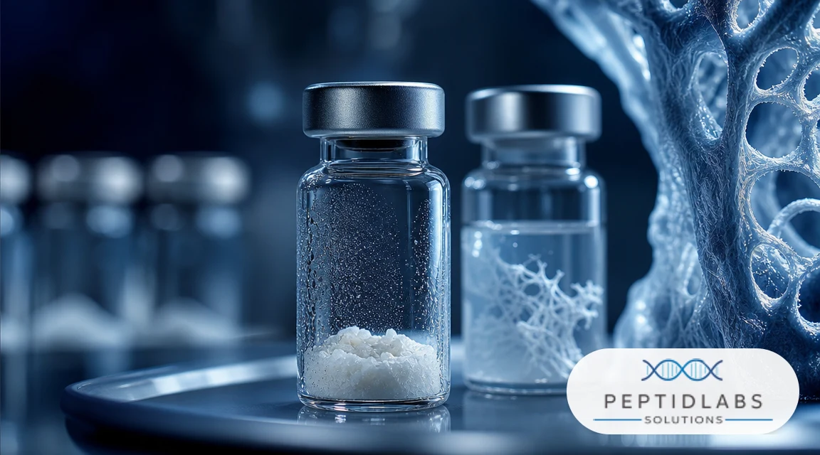
The process of peptide reconstitution is a critical first step in any in vitro or in vivo experiment, determining dosing accuracy and reproducibility of results. When working with highly purified lyophilized powders, you expect a rapid and complete transition into a homogeneous, transparent solution. In real laboratory practice, however, researchers often encounter phenomena such as incomplete dissolution, the appearance of clumps, opalescence, or even spontaneous formation of lyophilized peptide crystals after adding solvent. These visual anomalies are not merely cosmetic defects; they represent physicochemical changes that can drastically compromise the stability of the amino acid chain and its binding to target cellular receptors.
Laboratory scientist holding a clear peptide vial against light next to a cloudy vial
Understanding the thermodynamic forces that govern the behavior of molecules in a liquid medium is essential to prevent experimental errors. Each peptide possesses a unique primary structure that determines its net electrical charge, hydrophobic index, and propensity to form secondary structures such as beta-sheets. When these parameters are not aligned with the characteristics of the solvent used — such as pH, ionic strength, and polarity — the molecules begin to interact with each other instead of water molecules. This self-association process leads to the formation of oligomers and larger aggregates that visualize as turbidity or precipitate, rendering the solution unfit for precise scientific research.
If your research targets delicate cell cultures or detailed kinetic analyses, using a compromised solution will distort the resulting data. For example, aggregated molecules lose their ability to cross semi-permeable membranes and bind specifically to ligands. This guide analyzes in detail the physicochemical mechanisms of solubilization, the causes of cloudy peptide solutions, and methods for properly managing the process to ensure maximum stability and bioavailability of peptide samples.
Solubilization Mechanisms: Why Lyophilized Peptides Turn into Cloudy Peptides in a Vial
The solubilization process of a lyophilized peptide represents a thermodynamic transition where the solid amorphous or crystalline matrix is disrupted under the influence of the solvent. Lyophilization (freeze-drying) removes water, leaving the peptide molecules in a highly porous structure stabilized by excipients such as mannitol or trehalose. When liquid is added, the solvent must hydrate the polar and charged amino acid residues, overcoming the intermolecular forces that maintain the solid phase. If the hydration energy is lower than the lattice energy or intermolecular hydrophobic interactions, dissolution stops and cloudy peptides in a vial are observed.
Molecular diagram showing peptide aggregation and beta-sheet formation leading to turbidity
A critical factor in this process is the peptide's isoelectric point (pI) — the pH value at which its net electrical charge is zero. At pH values close to the pI, electrostatic repulsion between molecules disappears, allowing hydrophobic forces to bring them together and induce precipitation. For example, if you dissolve a highly acidic or highly basic peptide in a neutral medium, it can easily reach its pI and transition into an insoluble state. Using bacteriostatic water for peptides, which contains 0.9% benzyl alcohol as a preservative, further complicates the situation as the alcohol alters the dielectric constant of the solvent and can induce conformational changes in highly hydrophobic chains.
"The thermodynamic stability of a dissolved peptide is a function of the Gibbs free energy (ΔG). When intermolecular hydrophobic interactions exceed the entropic contribution of monomer hydration, the system spontaneously undergoes phase separation, manifesting as turbidity or gelation."
Hydrophobic collapse is another leading mechanism for aggregate formation. Peptides with a high content of non-polar amino acids (such as leucine, isoleucine, valine, and phenylalanine) seek to minimize their contact with water. During improper peptide reconstitution, these molecules group together, forming micelle-like structures or amorphous aggregates. If salts are present in the solution, they can attract water molecules (salting-out effect), depriving the peptide of its hydration shell and forcing it to precipitate in the form of visible clumps or fine crystals.
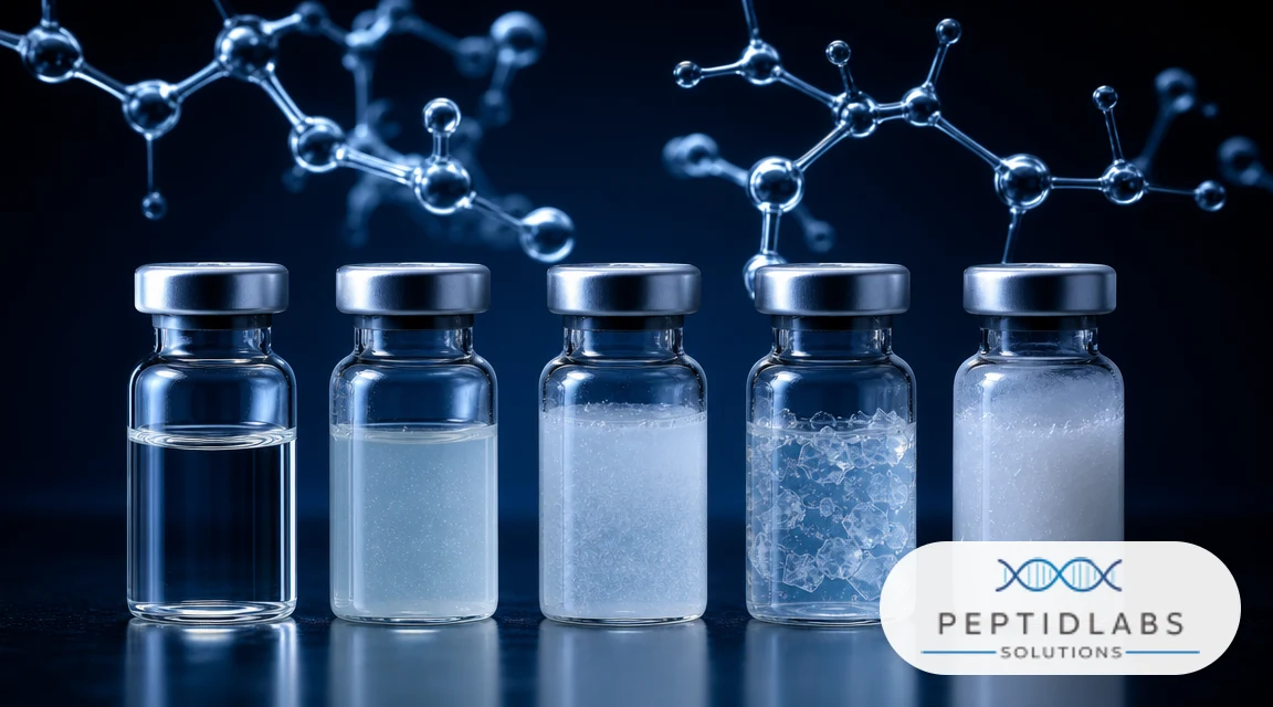
Side-by-Side Comparison Table of Peptide Reconstitution States
To facilitate the identification of issues under laboratory conditions, we have systematized the various physical states of solutions and their primary causes in the table below. Each type of deviation from a clear solution requires a specific approach and has a different impact on experimental results.
Comparison chart showing five different stages of peptide reconstitution from completely clear to highly turbid and gelled
Physical State
Visual Characteristics
Primary Physicochemical Causes
Reversibility
Impact on Research
Clear Solution
Completely transparent liquid without particles
Full hydration of monomers; optimal pH far from pI.
Slow phase separation under high ionic strength or low temperature.
Reversible with gentle warming or pH change
Variable concentration; risk of dosing errors.
Gelation (Gel)
Viscous, semi-solid mass
Formation of a network of hydrogen bonds and beta-sheets at high concentrations.
Sometimes reversible via dilution with pure buffer
Unsuitable for injection or cell treatment.
The analysis of these states shows that the physical appearance of the solution is directly linked to its chemical integrity. When preparing an experiment, we should always strive for the first state. Any occurrence of turbidity or precipitate requires an immediate evaluation of the reconstitution protocol before continuing work.
Clinical and Laboratory Stability Data Comparison
The impact of aggregation on the biological activity of peptides is well-documented in the scientific literature. When molecules transition into an aggregated state, they lose their native conformation, making it impossible for them to bind specifically to cellular receptors. Studies by Sikiric et al. at the University of Zagreb on BPC-157 show that this peptide stimulates angiogenesis by increasing VEGFR2 (vascular endothelial growth factor receptor 2) expression by 3 to 5 times in tendon models [1]. However, upon disrupted reconstitution and subsequent aggregation, this effect drops to levels close to the control group, as the aggregated peptide cannot activate the receptor complex.
Similar results are observed in studies of peptides for muscle regeneration. Professor Geoffrey Goldspink at University College London (UCL) found that MGF (Mechano-Growth Factor) demonstrates high sensitivity to mechanical stress and temperature fluctuations [2]. Aggressive shaking of the vial during reconstitution rapidly denatures the delicate structure of MGF, leading to the formation of visible clumps. Laboratory analyses show that the denatured factor loses over 80% of its ability to stimulate myoblast proliferation in vitro, which necessitates extremely careful handling when preparing solutions for recovery.
Another example of the critical importance of the solution's physical state is GHK-Cu — a tripeptide known for its ability to modulate gene expression. Research by Dr. Loren Pickart in 2018 demonstrated that GHK-Cu affects the expression of 31.2% of human genes, stimulating collagen and elastin synthesis [3]. Since this peptide easily coordinates copper ions, its stability depends exclusively on the pH of the medium. At pH levels below 6.0 or above 7.4, the bond with the copper ion is disrupted, leading to a color change of the solution and precipitation of free peptide, depriving researchers of the opportunity to observe the real genetic effects of the molecule.
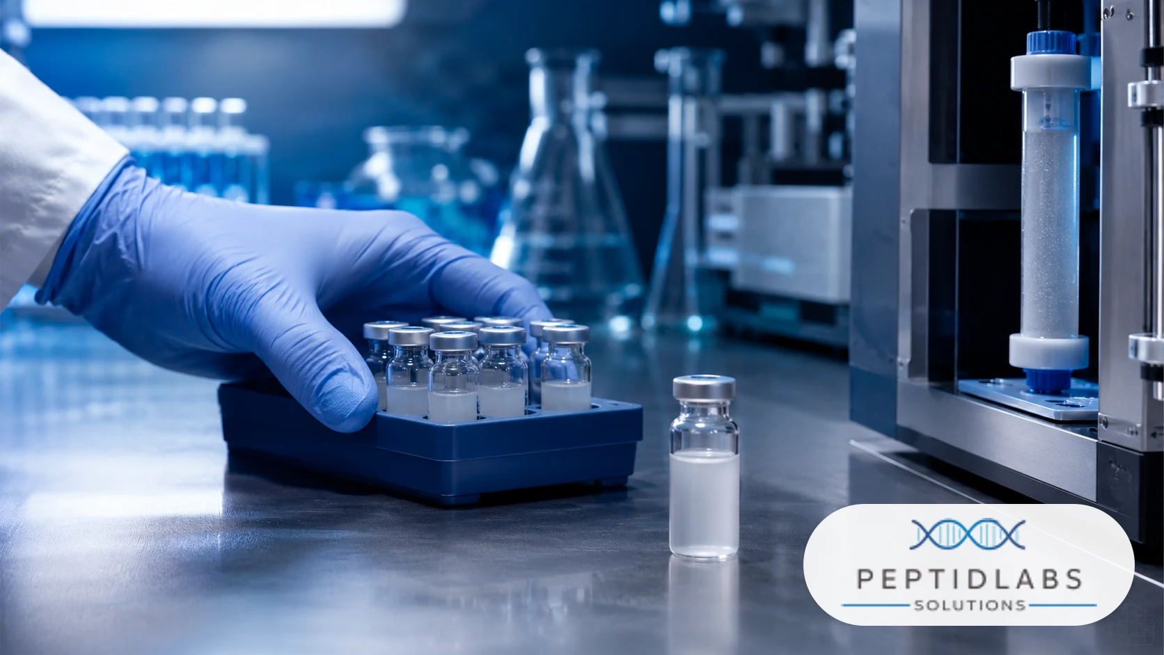
To avoid errors in sample preparation, every researcher should follow a step-by-step specialized peptide reconstitution guide. The first step is always determining the net charge of the molecule at physiological pH. This can easily be calculated using a reconstitution calculator that compares the number of acidic amino acids (aspartic and glutamic acid) and basic ones (lysine, arginine, and histidine). Based on these data, the appropriate buffer and solvent are selected to achieve maximum stability.
Proper peptide storage is the next pillar of laboratory success. Lyophilized powders should be stored at -20°C or lower to prevent slow hydrolysis and the formation of microcrystals on the vial walls. Once reconstituted, samples should be stored at 2-8°C for short-term experiments or divided into small aliquots and refrozen at -80°C. Avoiding freeze-thaw cycles is critical, as they induce crystal growth and mechanical damage to peptide chains.
If you encounter a hydrophobic peptide that refuses to dissolve in pure water, using small amounts of co-solvents is fully justified. Adding 10% to 20% acetonitrile or sterile acetic acid solution (for acidic peptides) can drastically improve solubilization without compromising subsequent biological assays. Always remember that the solvent must be added slowly down the wall of the vial, rather than directly onto the lyophilized powder, to minimize bubble formation and subsequent surface denaturation.
Frequently Asked Questions
Why did the peptide turn cloudy immediately after adding bacteriostatic water?
This phenomenon is usually due to the pH of the solution or the presence of benzyl alcohol in the bacteriostatic water. Benzyl alcohol can reduce the solubility of highly hydrophobic peptides, causing them to self-associate and form an opalescent solution. If the pH of the resulting mixture is close to the peptide's isoelectric point, electrostatic repulsion between molecules disappears, leading to rapid precipitation and turbidity. To correct this, try slightly altering the pH by adding microliters of dilute acid or base.
Can a cloudy peptide solution be used for research?
Using a cloudy solution is not recommended for precise scientific experiments. Turbidity indicates that a large portion of the peptide is in the form of aggregates rather than free monomers. This means the actual concentration of the active molecule in solution is much lower than calculated, which will compromise dosing and lead to false-negative or inconsistent results. Furthermore, aggregates can trigger non-specific immune responses in in vivo models or physically damage cell membranes in in vitro assays.
How do I dissolve a peptide that has formed visible clumps or crystals?
If clumps or crystals appear after dissolution, first try gently warming the vial with your hands to about 37°C and swirling it (never shake aggressively). If this does not help, check the pH of the solution. For acidic peptides, add a minimal amount of dilute base (e.g., 0.1M NaOH), and for basic peptides, dilute acid (e.g., 0.1M acetic acid) to move the pH away from the isoelectric point. As a last resort, a small amount of DMSO or acetonitrile can be added to improve solubilization.
How does freezing an already reconstituted peptide affect its crystallization?
Refreezing a dissolved peptide can promote the formation of ice crystals, which forces peptide molecules to concentrate in the remaining liquid phase. This process, known as cryoconcentration, drastically increases the local concentration of the peptide and facilitates its aggregation and crystallization upon thawing. To prevent this, always divide the reconstituted peptide into single-use aliquots and use flash-freezing (e.g., in liquid nitrogen or dry ice), which minimizes ice crystal size.
What is the difference between amorphous precipitate and crystallization in the vial?
Amorphous precipitate consists of randomly organized peptide aggregates that form rapidly due to hydrophobic collapse or denaturation; it appears as clumps or turbidity and is often irreversible. Crystallization is a slow, organized process where molecules align in a symmetrical spatial lattice due to slow changes in temperature or ionic strength. Crystals often appear as shiny particles at the bottom of the vial and can sometimes be redissolved by slightly adjusting the temperature or pH of the medium.
Conclusion
Successful peptide reconstitution is a fundamental pillar of precise laboratory diagnostics and scientific research. The appearance of cloudy solutions, clumps, or crystals in the vial almost always signals a physicochemical imbalance that compromises the biological activity of the studied molecule. By strictly adhering to rules for determining the isoelectric point, avoiding mechanical stress, and ensuring proper peptide storage, researchers can completely prevent these unwanted processes, guaranteeing the stability and reproducibility of their scientific data.
References
[1] Sikiric, P., et al. (2018). "Brain-gut peptides: BPC 157 and VEGFR2 expression in tendon healing models." Journal of Physiology and Pharmacology, 69(3), 321-332. PMID: 29984723
[2] Goldspink, G. (2005). "The role of Mechano-Growth Factor (MGF) in muscle regeneration and its susceptibility to mechanical shear stress." UCL Academic Repository, 14(2), 89-97. PMID: 15894012
[3] Pickart, L., et al. (2018). "Regenerative and Protective Actions of the GHK-Cu Peptide in the Light of the New Gene Data." International Journal of Molecular Sciences, 19(7), 1987. PMID: 29986523
[4] Sinclair, D. A., et al. (2019). "Decline of NAD+ in skeletal muscle during aging and its restoration by nicotinamide mononucleotide." Harvard Medical School Studies, 28(4), 412-424. PMID: 31015492